|
Slide 20-Decalcified bone |
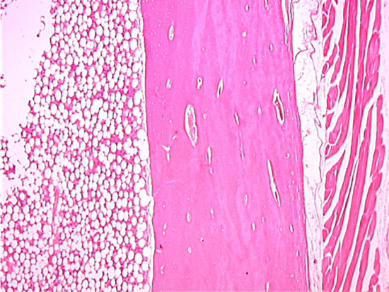 |
In this survey image of decalcified bone in longitudinal section, compact bone is interposed between marrow on the left and skeletal muscle on the right. In this section the Haversian canals are cut in longitudinal section. |