|
Slide 94-Human duodenum |
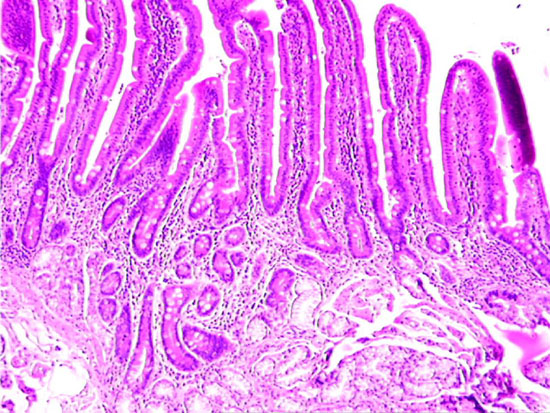 |
In this image, finger-like villi and intestinal glands can be seen. Note the pale-staining goblet cells interspersed throughout the absorptive epithelium, the loose connective tissue (lamina propria) that forms the core of each villus, and the intestinal glands in the lamina propria beneath the villi. At bottom, a portion of Brunners gland can be seen. |