Musculo-Skeletal Examination
Hand and Wrist
Normal function of the hand and wrist is obviously of great importance. A cursory review of this area is included in the Upper Extremity Examination. What follows is a description of commonly occurring pain syndromes and pathologic processes involving this region.
For Additional Information See: Digital DDx: Hand Pain
- Carpal Tunnel Syndrome
Presentation and Anatomy: The median nerve travels through a narrow space when it crosses the wrist en route to the hand. Occasionally, this space becomes inadequate to accommodate the nerve, placing it under increased pressure. The precise reason why this occurs is not clear. Patients usually report some combination of the following:
- Numbness and tingling (ie neuropathic pain symptoms) in the distribution of the median nerve (thumb, index, middle and lateral 1/2 of ring finger)
- Symptoms are often worse at night, presumably due to tendency to flex wrist during sleep. Flexing puts additional pressure on the nerve.
- Patients will often try to "shake out" their hands in an effort to reduce pain and "increase blood flow" (based on the patient's assumption that decreased perfusion caused the symptoms).
- With increased severity, pain can be present at all times during the day.
- In severe cases, there may be loss of motor strength of the thumb.
Examination:
- The hand and wrist usually appear normal
- Pain may some times be reproducible by tapping over the nerve (Tinnel's
sign). It may also occasionally be reproducible if the wrist is held in
forced flexion x 1 minute (Phelan's sign). Neither of these signs is particularly
sensitive.
 Tinnel's Test
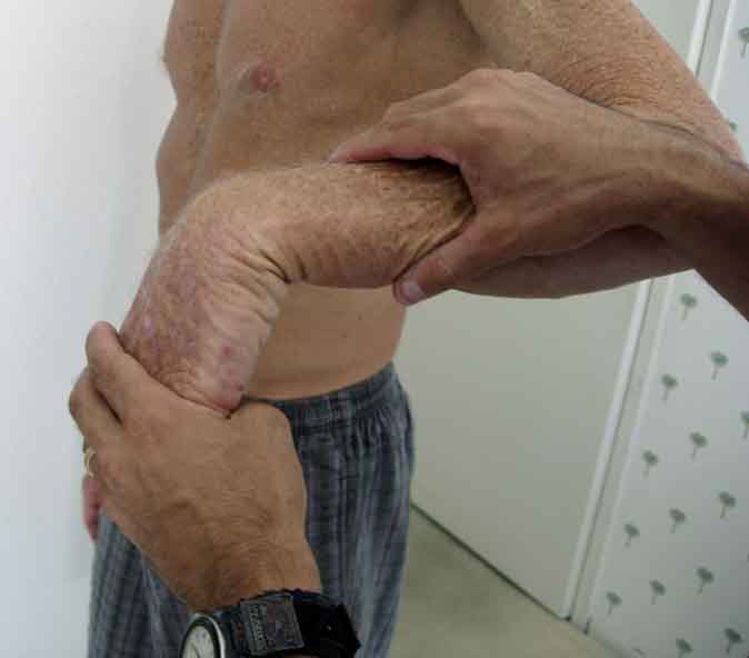 Phelan's Test
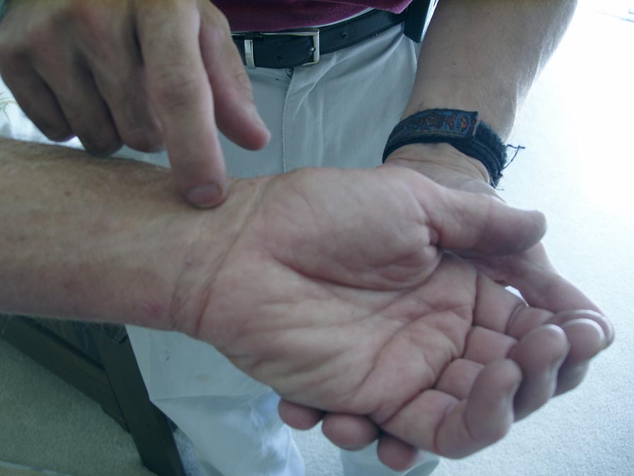
 - In advanced carpal tunnel, there may be atrophy of the thenar eminence
(due to denervation of the muscle as well as disuse atrophy) and associated
decrease in motor strength. The Abductor Pollicis Brevis (APB) muscle
receives sole innervation from the median nerve. Function can be tested
by providing resistance to abduction up and away from the plane of the
palm.
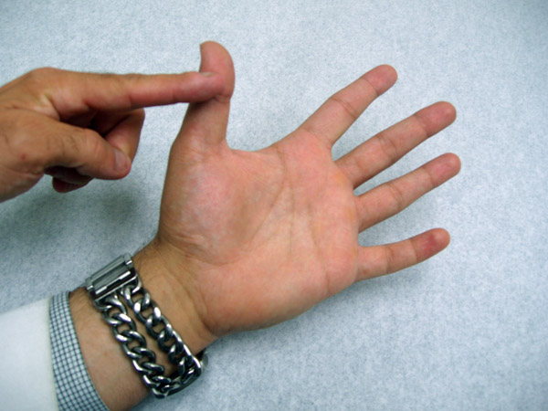 Resistance to movement of APB
 - Prolonged compression will lead to impaired 2 point discrimination on sensory
testing. That is, the patient can't discern whether being touched with one object
or 2 when separated by 5mm (can check using a bent paper clip).
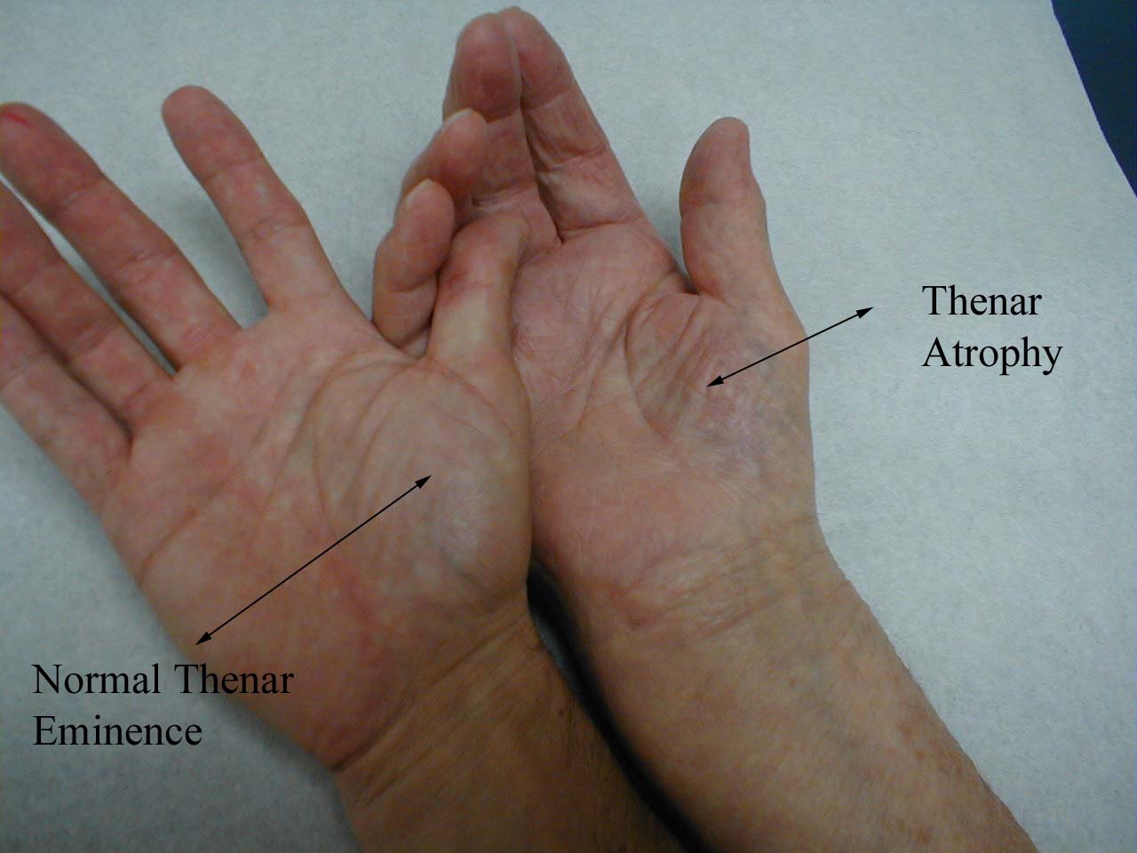 Carpal Tunnel Induced Atrophy:Chronic, severe compression of the median nerve within the carpal tunnel has led to atrophy of the Thenar muscles (hand on right). A normal appearing Thenar Eminence is demonstrated on left.
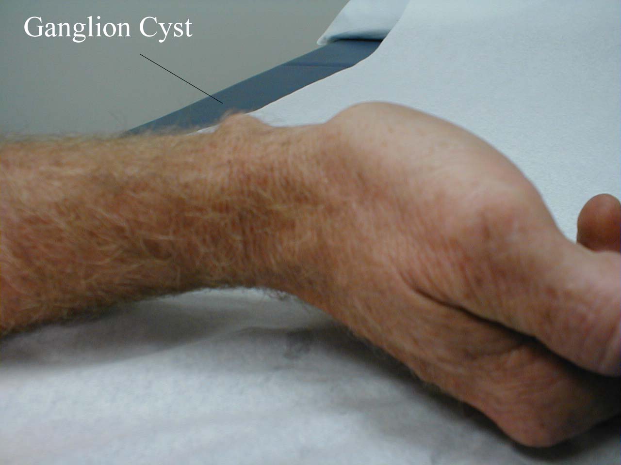
- Ganglion Cyst
Presentation and Anatomy: Idiopathic, spontaneous protrusion of joint fluid outside of the articular space. The cyst is painless and usually located on the dorsal aspect of the wrist.
Examination:
- Patients often present noting the abrupt development of a focally swollen area.
- There is usually no associated pain or inflammation.
- On palpation, the structure has a fluid filled consistency and is non-tender.
- The structure should trans-illuminate when a light is placed upon it (as it's fluid filled).
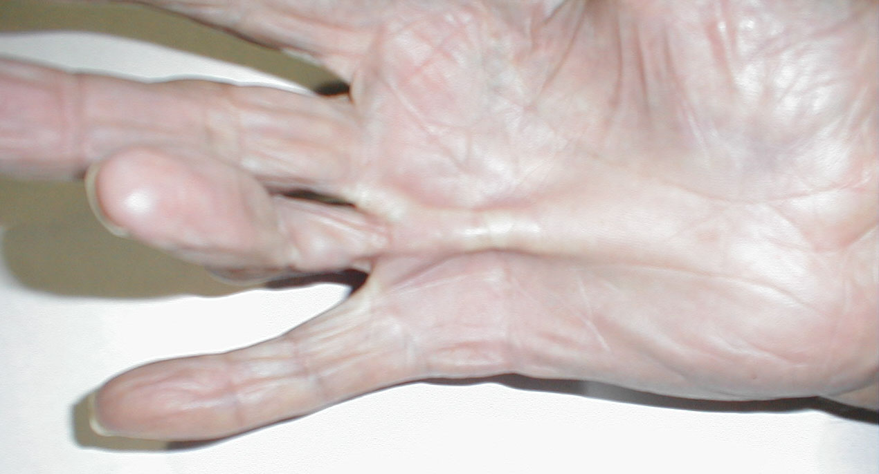
- Dupuytren's Contracture Presentation and Anatomy: Thickening of the
palmar fascia, which is usually painless and develops slowly over time. If
pronounced, it may prevent the hand from being able to fully open.
Examination:
- Obvious focal thickening on palmar aspect hand.
- On palpation, feels tough and thick, though non-tender and without signs of inflammation.
- May interfere with ability to fully open hand
Dupuytren's Contracture
- Heberden's Nodes
Presentation and Anatomy: Bony excresences that cause deformity at the DIP joints of the fingers. Occurs slowly over time and is associated with Osteoarthritis. May affect many joints or only a few, though not usually symmetric. Similar protrusions at the PIP joints are called Bouchard's nodes.
Examination:
- Obvious bony protrusions at DIP joints
- Non-tender on palpation with an absence of inflammation
- Some times interfere with joint movement and function
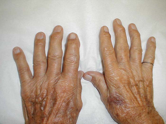 Heberden's Nodes
- Trigger Finger
Presentation and Anatomy: Flexor tendons connect muscles proximal to the wrist to the fingers. When the muscles shorten, they pull on the tendons, causing the fingers to flex. Occasionally, nodules/irregularities develop along the tendons, which then interfere with their smooth movement thru "pulleys" on the palm. Patients note difficulty flexing and extending the affected finger and lack of smooth movement. This is associated with a sensation of sudden freeing of the tendon ("triggering") when the irregularity slips through the pulley.
Examination:
- The palm and fingers usually appear normal. The affected tendon is not visible.
- Ask the patient to fully flex the affected finger. When they attempt to extend and flex it, the movement will be impaired. It's worth noting that sometimes the triggering does not occur with every movement.
- If you place one of your fingers over the affected tendon, you may feel the "pop" when it finally pulls thru. There is usually no associated pain or inflammation.
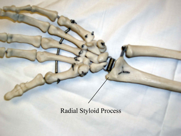
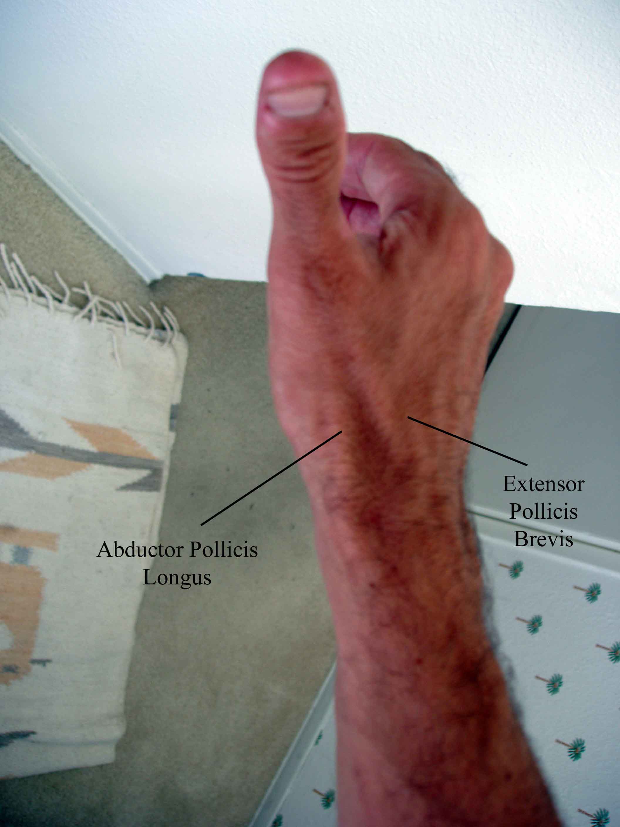
- Tenosynovitis of the Thumb (DeQuervain's type)
Presentation and Anatomy: Repetitive abduction and adduction of the thumb can irritate the tendons of the extensor policis brevis and abductor policis longus muscles. When this occurs, any movement of the thumb (in particular, gripping) may cause pain at its base.
Examination:
- The thumb usually appears normal. In cases of severe tendonitis, there may be swelling overlying the tendons.
- Tenderness at the point where the tendons of the extensor pollicis brevis
and longus cross the radial styloid (distal end of the radius)
- Pain with passive stretching of the tendons (a.k.a. Finklestein Test):
- Direct the patient to place the thumb in their palm.
- Have them cover the thumb with the fingers of the same hand, forming a fist.
- Gently deviate the wrist towards the ulna. This stretches the inflamed tendons over the radial styloid, reproducing the patient's pain.
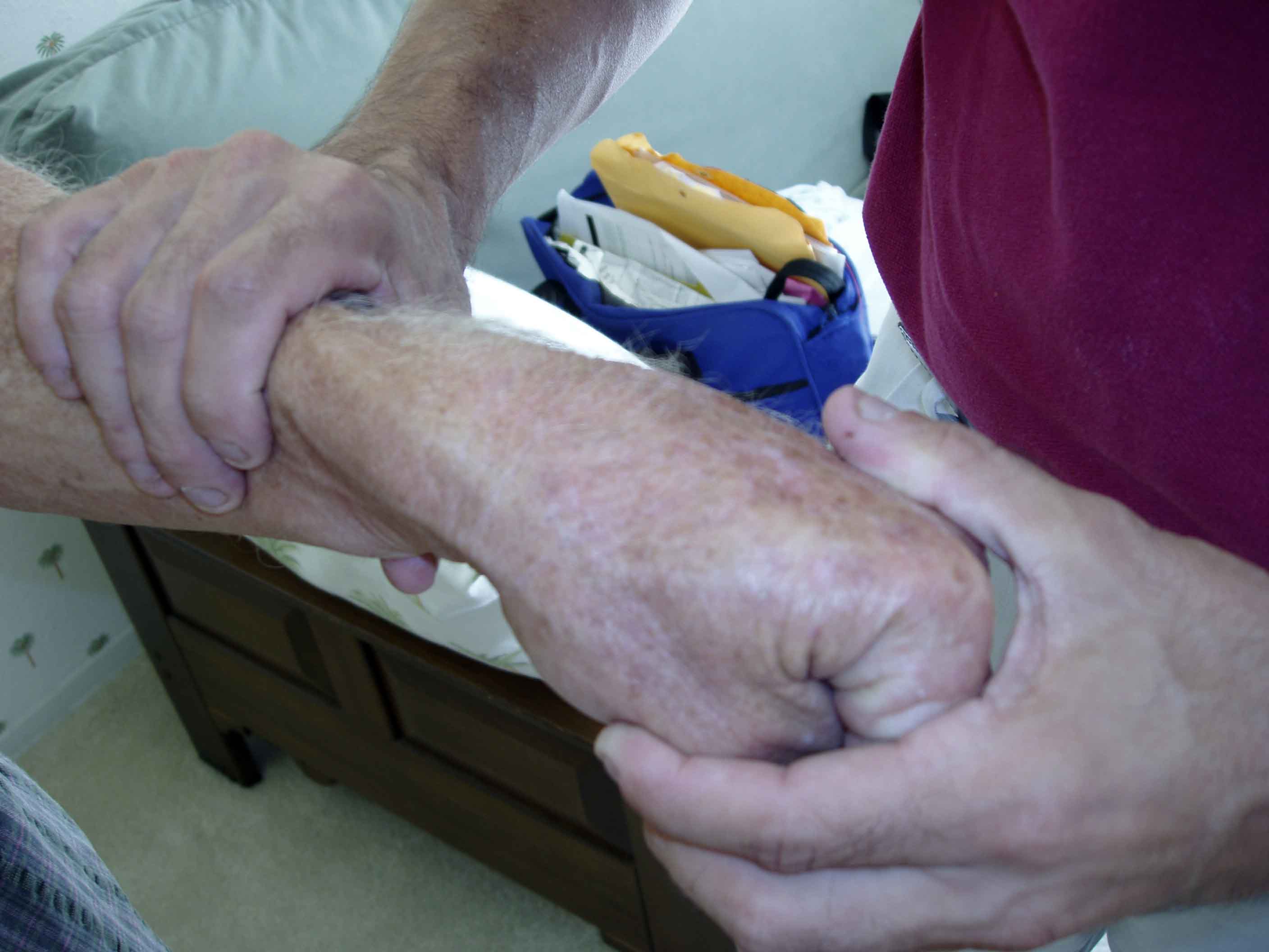 Finkelstein's Test
Selected Traumatic Injuries To The Hand (not in any way inclusive!)
- Boxer's Fracture
Presentation: When a closed fist strikes a solid surface, the force may cause a break in the 5th metacarpal.Examination:
- Pain and swelling over the 5th metacarpal
Pathoantomy of Boxer's Fracture
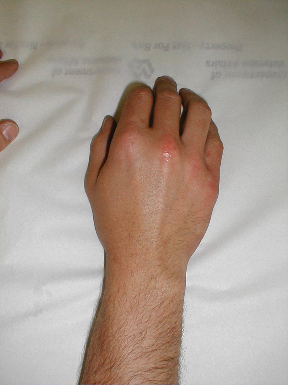
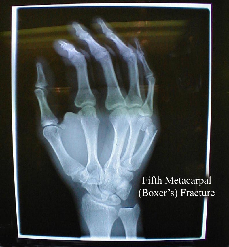 Boxer's Fracture. Note swelling over dorsal aspect of right hand, most pronounced below the small finger. It is the result of a fracture to the fifth metacarpal (bone below small finger), usually associated with striking an object with a closed fist. X-ray demonstrates fracture of distal right metacarpal.
- Pain and swelling over the 5th metacarpal
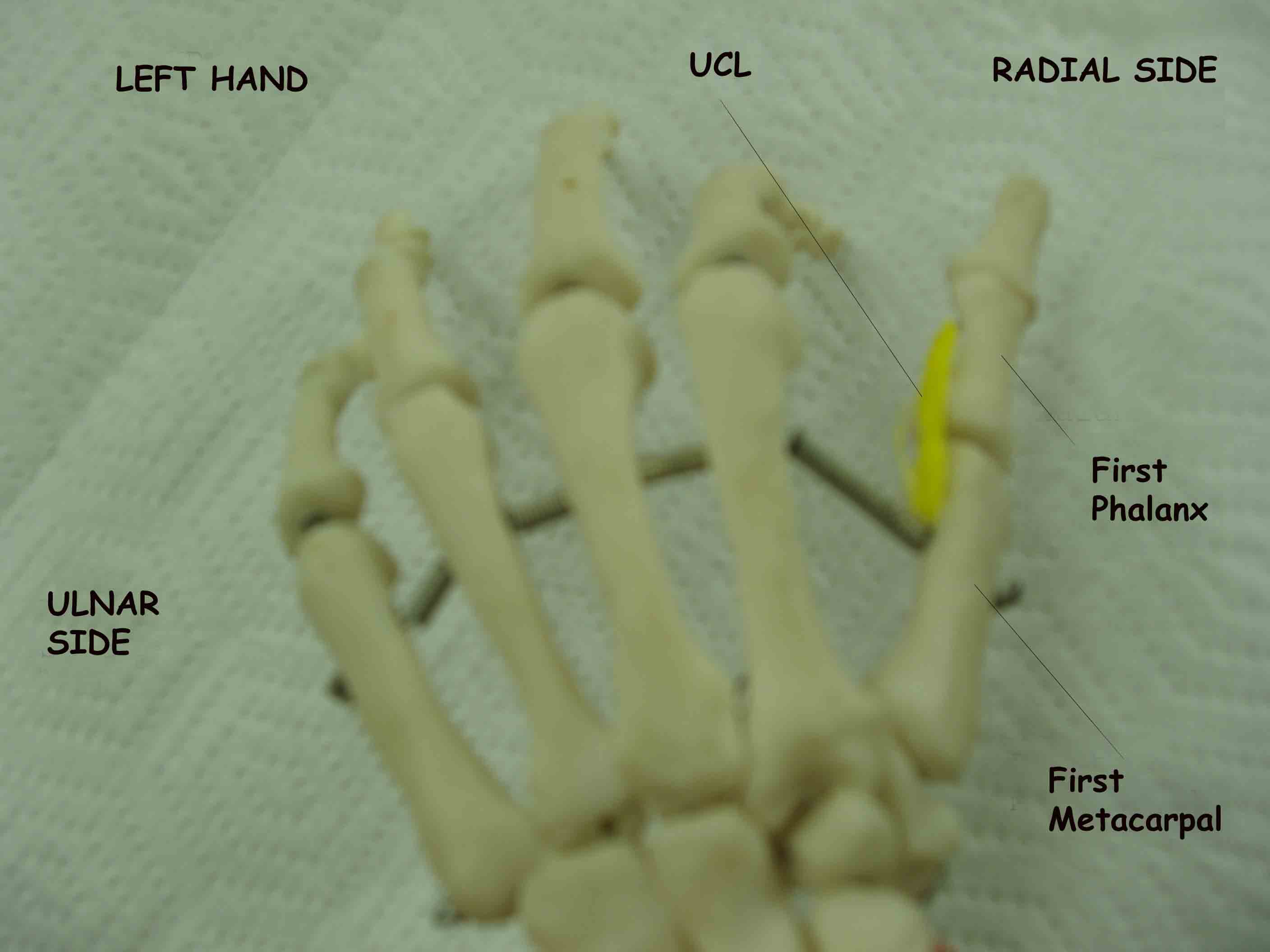
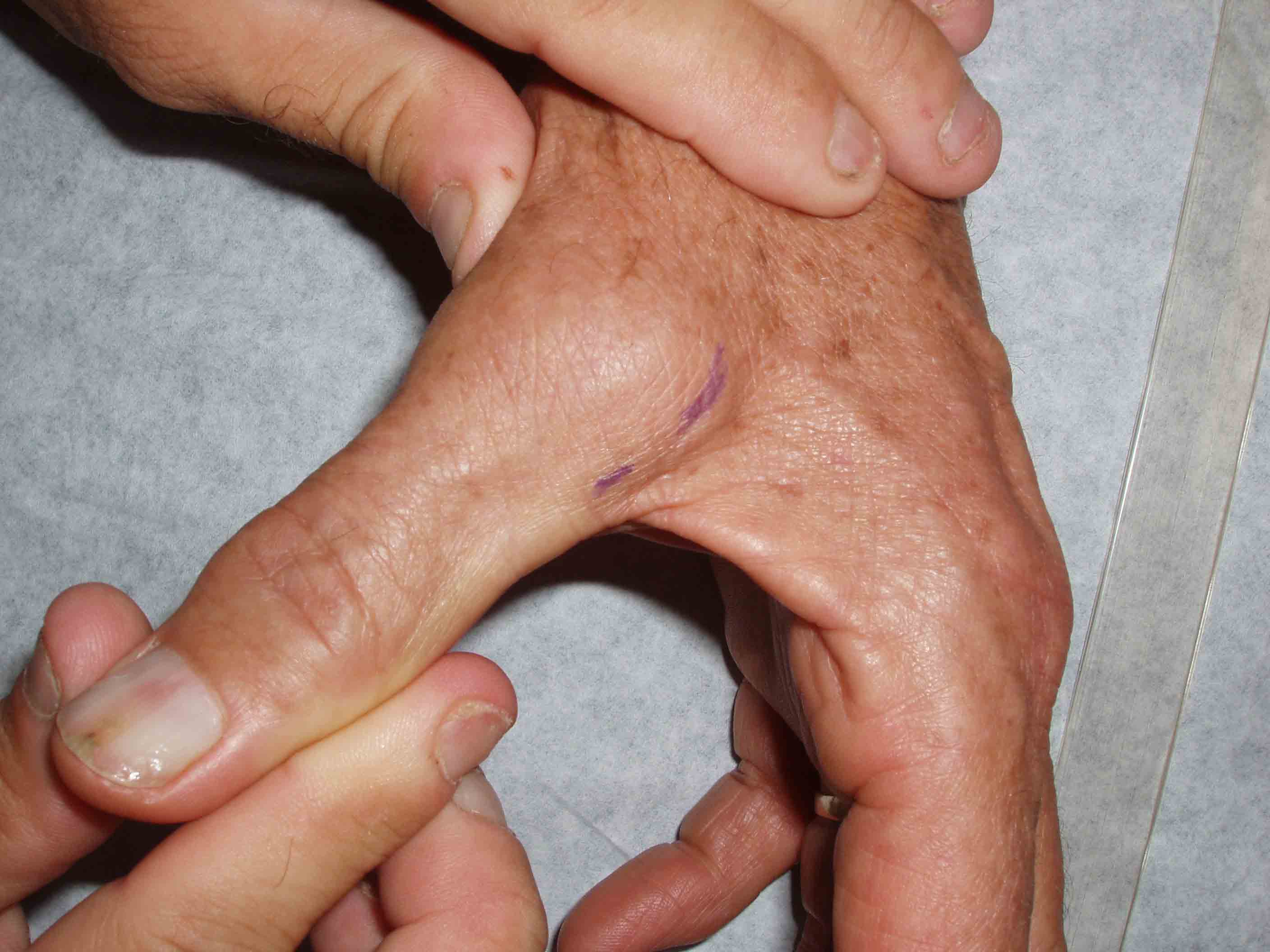
- Ulnar Collateral Ligament Disruption (gamekeeper's Thumb)
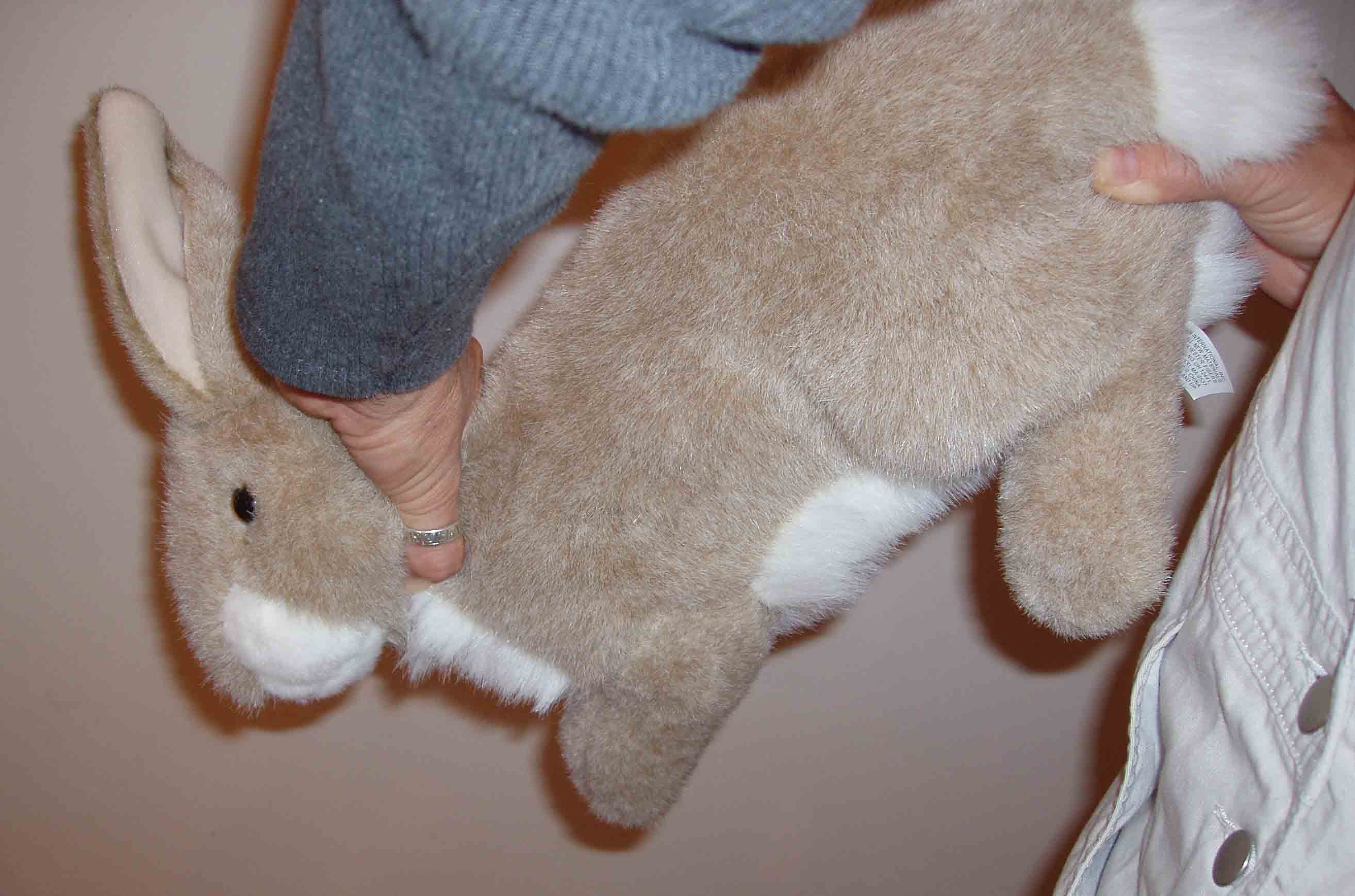
(Don't Worry, Bunny Used In Photo Is Not Real!)
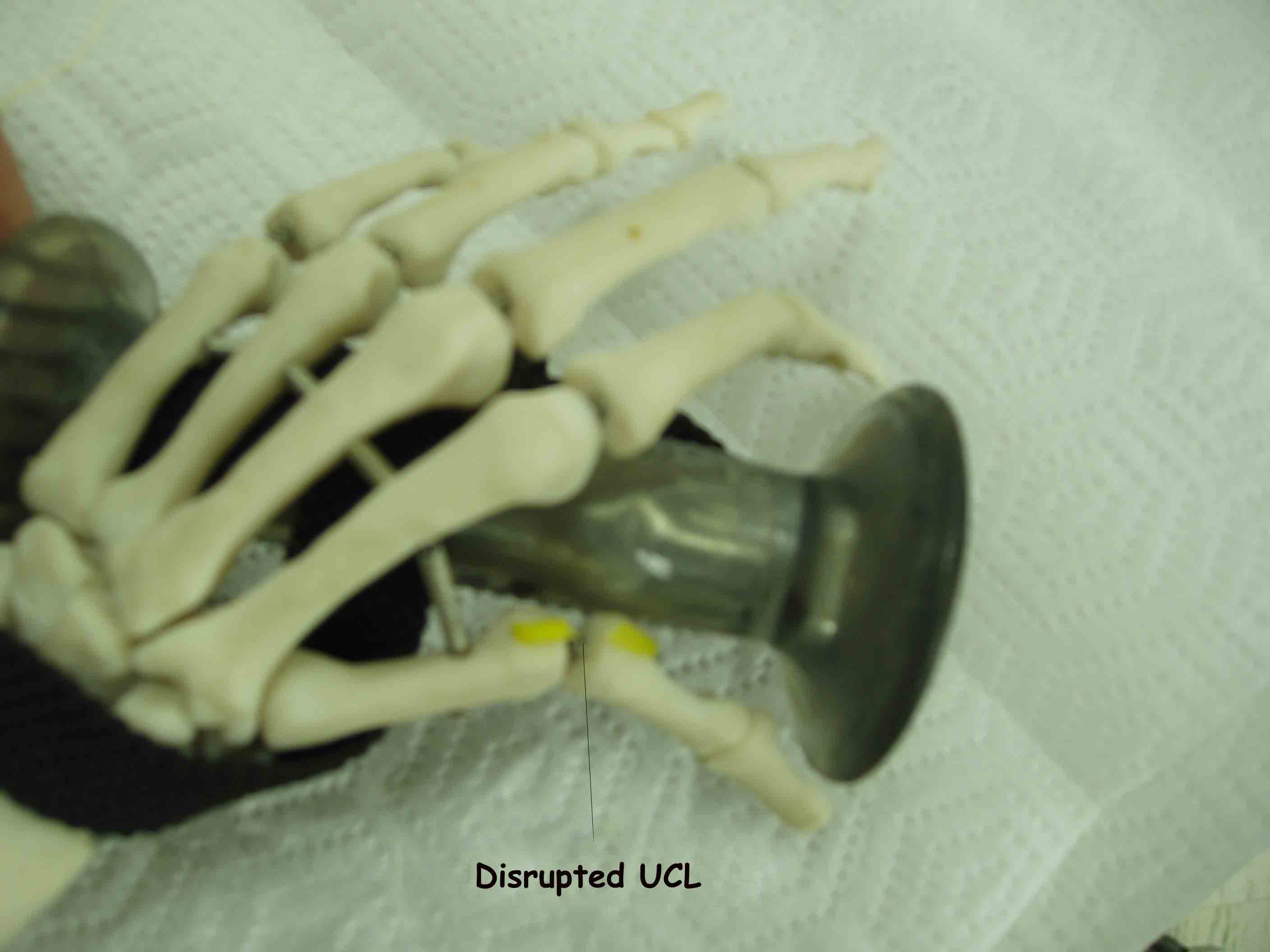
The ulnar collateral ligament (UCL) is a strong band of tissue that connects the first phalanx of the thumb to the metacarpal bone along the ulnar side. Injury to this structure was first described in Scottish gamekeepers, who damaged the ligament as a result of the manner in which they killed rabbits. The head of the rabbit was grasped between thumb and first finger of one hand while they pulled on the rabbit's hind quarters with their other. This force chronically stressed the UCL, leading to weakening or frank rupture. After its initial description, it was quickly recognized that the ligament could be torn by any strong force that acutely abducts the extended thumb. Patient's are usually immediately aware that something is wrong, developing swelling, pain and instability at the metacarpal-phalangeal (MCP) joint . It has become a relatively common ski injury, occurring when a person falls on a hand that has a ski pole gripped between the thumb and forefinger.
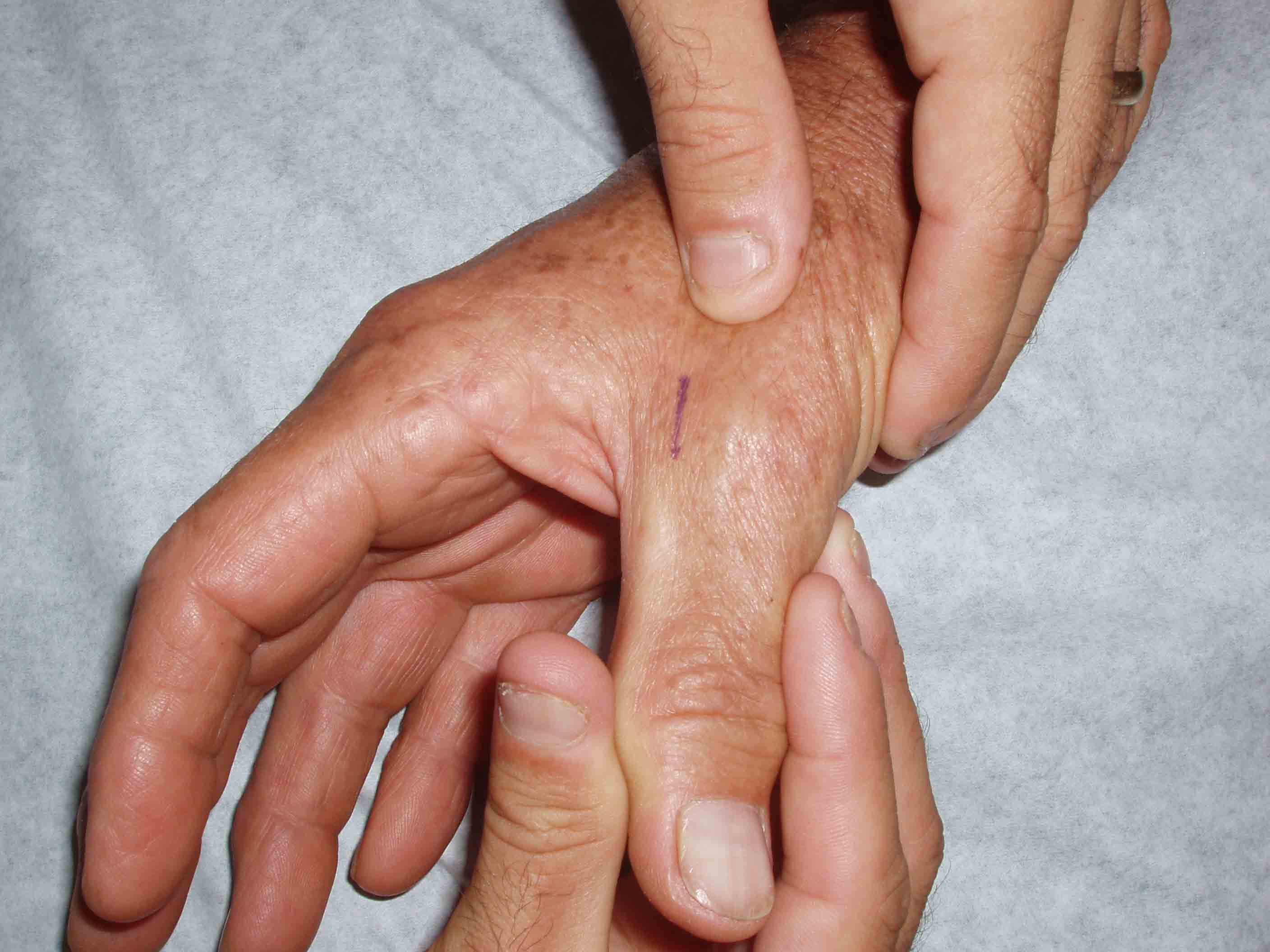
Examination is remarkable for swelling and pain at the MCP. The key maneuver assesses the degree of laxity at the joint. Place the thumb in extension (see picture below for positioning). Gently grasp the end of the thumb and apply an abducting force. If the UCL has been disrupted, you will be able to distract the thumb to a much larger degree then when compared to the normal side.
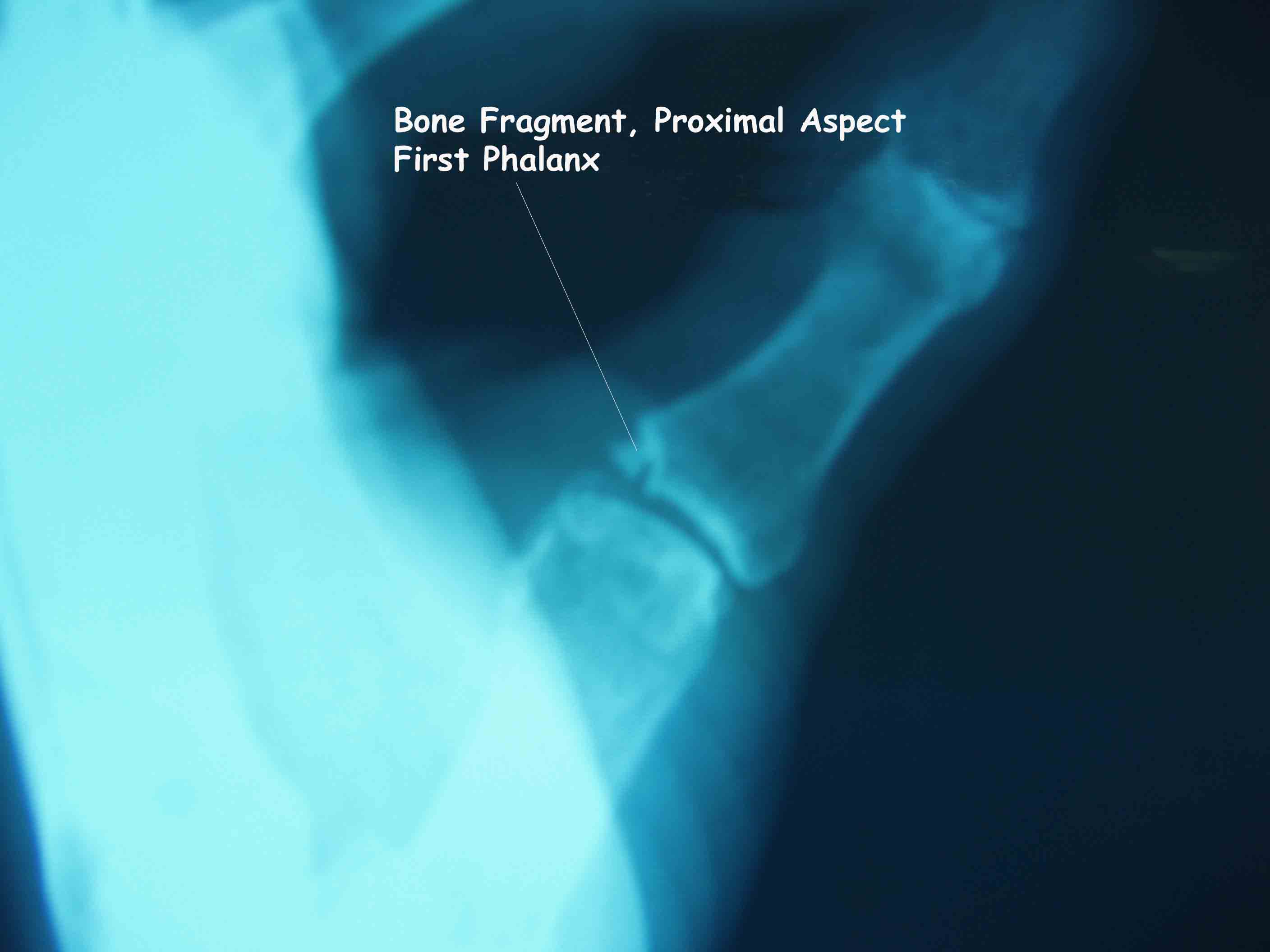
The ligament may become disrupted at it's insertion on the proximal phalanx, pulling away a small piece of bone that can be seen on x-ray.